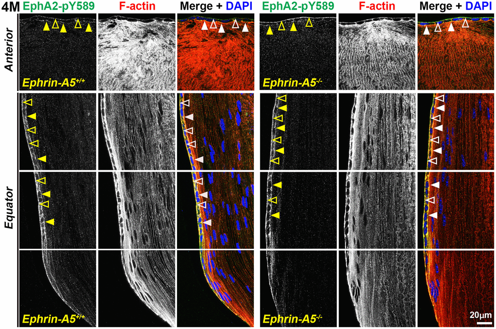
Figure 6.Canonically active EphA2-pY589 protein is enriched at basal and apical membranes of lens epithelial cells. Images of the equator region were taken in sequence along similar areas of the lens. In 4-month-old control and ephrin-A5-/- longitudinal lens sections stained with EphA2-pY589 (green) antibody, phalloidin (F-actin, red), and DAPI (nuclei, blue), the EphA2-pY589 signal was visible in anterior lens epithelial cells with enrichment at the basal (open arrowheads) and apical (arrowheads) membranes. There was increased EphA2-pY589 staining signal in equatorial epithelial cells with enrichment at the basal (open arrowheads) and apical (arrowheads) membranes. EphA2-pY589 staining was also visible between equatorial epithelial cells along the lateral membranes. There are no obvious differences in staining signal and localization between control and ephrin-A5-/- sections. Scale bar, 20 μm.