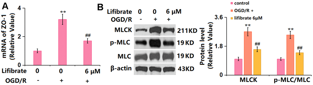
Figure 7.Lifibrate inhibited activation of the MLCK/p-MLC signaling against OGD/R. Cells were exposed to hypoxic conditions for 6 h, followed by exposure to reperfusion media for 24 h in the presence or absence of Lifibrate (6 μM). (A) mRNA of MLCK; (B) Protein levels of MLCK and p-MLC/MLC (n=6, **, P<0.01 vs. vehicle group; ##, P< 0.01 vs. OGD/R group).