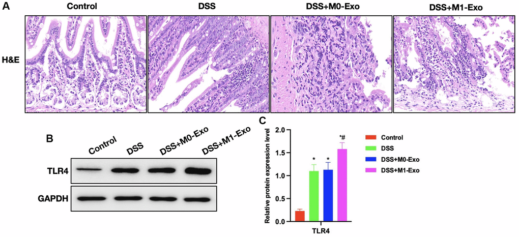
Figure 3.Role of M1-Exo in the pathology of colitis. (A) H&E (n = 5). Intestinal tissue in DSS group showed apparent edema, inflammatory tissue, and epithelial cell injury, while that in M1-Exo group showed aggravated mucosal destruction and obvious inflammatory response. (B, C) Relative protein levels (n = 5). TLR4 of DSS group were up-regulated, and further increased in M1-Exo group. *P < 0.05 versus Control group, #P < 0.05 versus DSS group.