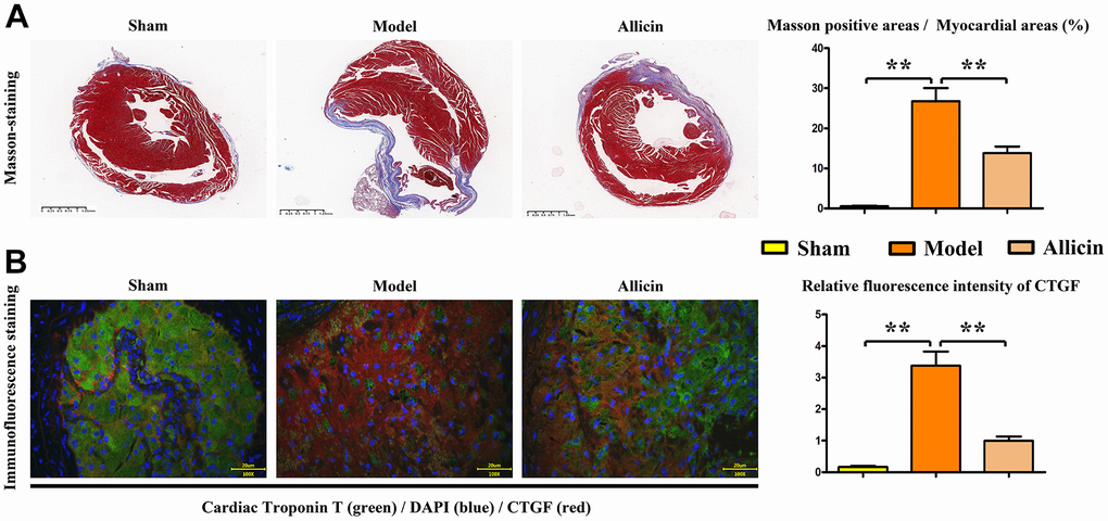
Figure 3.Quantification of myocardial fibers and immunofluorescence staining of CTGF in Masson's-stained mice. (A) Masson's staining of mouse hearts and the corresponding data. (B) Dual immunofluorescence staining of myocardial troponin T (T) and CTGF, and statistical analysis of CTGF intensity to evaluate the severity of cardiac fibrosis. One-way analysis, **p < 0.01, n = 6/group.