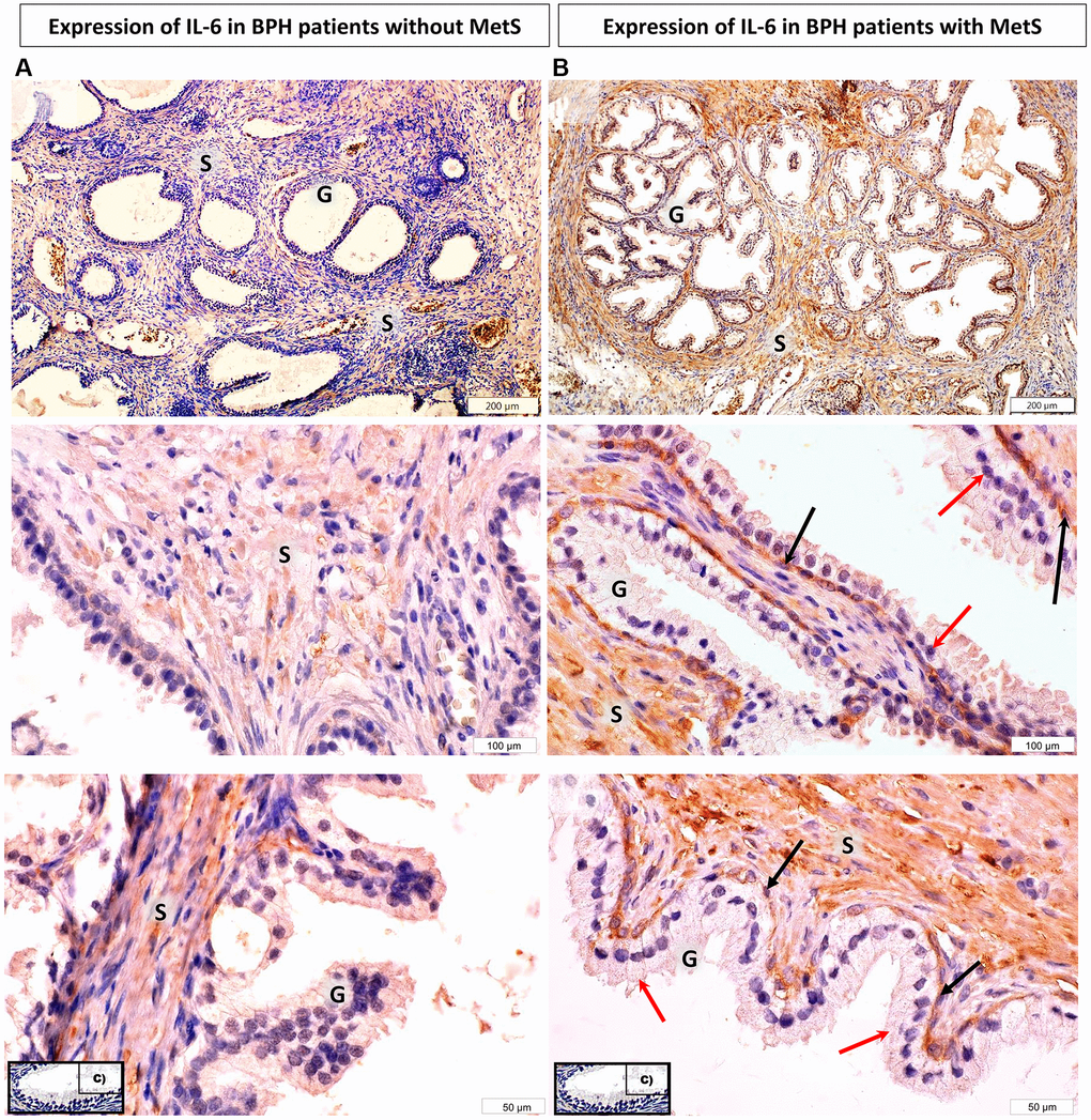
Figure 1.Microscopic image of the expression of the cytoplasmic immunohistochemical reaction to IL-6. (A) microscopic specimen of the prostate in a patient with benign prostatic hyperplasia and without MetS, (B) microscopic specimen of the prostate in a patient with benign prostatic hyperplasia and MetS. C - negative control (reaction without the use of an antibody). The cytoplasmic immunohistochemical reaction expressing IL-6 was stained brown (DAB +), S - prostate stroma, G - glandular part, ↑ - luminal cells, ↑ basal cells.