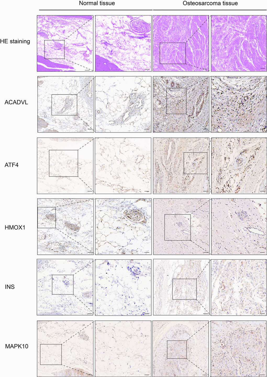
Figure 5.Hematoxylin eosin (HE) staining and immunohistochemistry (IHC) staining of normal and osteosarcoma tissue. Scale bar, 100 μm (left panel) and 50 μm (right panel).
Figure 5 — Construction and validation of an oxidative-stress-related risk model for predicting the prognosis of osteosarcoma | Aging