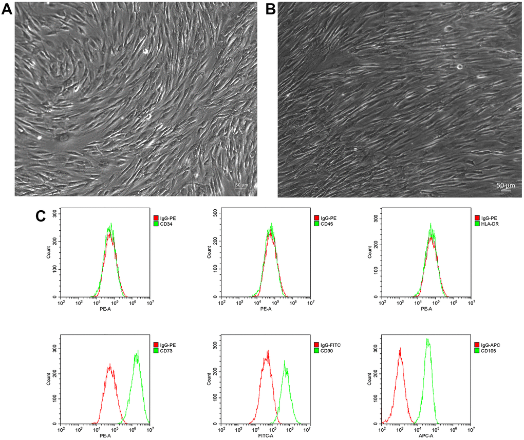
Figure 1.Isolation and phenotyping of in vitro cultured NP-MSCs. Representative phase-contrast images of passage 1 (A) and passage 2 (3 weeks) (bar = 50 μm) (B) NP-MSCs showing adherent cells with a long-spindle shape morphology. (C) Analyses of cell surface markers of NP-MSCs by flow cytometry, showing positive CD105, CD90, CD73 and negative CD34, CD45 and HLA-DR. All the samples were from case 5.