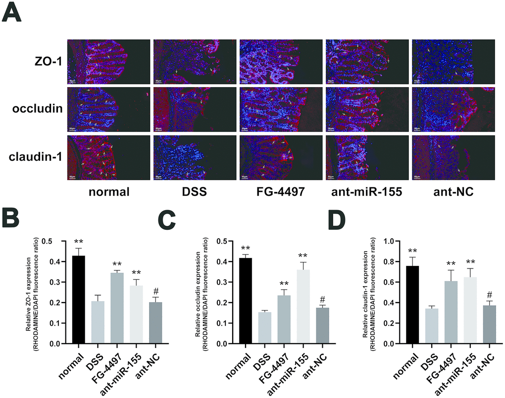
Figure 5.Immunofluorescence assay presented the expression of TJ proteins in mice colon. (A) Immunoflorescent staining of ZO-1, occludin, and claudin-1 in the colonic tissues. (magnification, ×200) Red signal (rhodamine) labeled by green arrows was positive for these proteins. DAPI (blue) was used to counterstain of nuclei. (B–D) Relative ZO-1, occludin, and claudin-1 expression measured by rhodamine and DAPI fluorescence ratio in all groups were presented. Each bar represents mean ± SD, n=5 from each group, #P > 0.05, *P < 0.05, **P < 0.01 vs. DSS group.