Submit an Article
Navigate
Home
Editorial Board
Editorial Policies
Current Volume
Archive
Scientific Integrity
Publication Ethics Statements
Interviews with Outstanding Authors
Newsroom
Sponsored Conferences
Podcast
Contact
Special Collections
Submit an Article
Online ISSN: 1945-4589
Research Paper
|
Volume 12, Issue 13
|
pp. 13647–13667
MiR-146b-5p suppresses the malignancy of GSC/MSC fusion cells by targeting SMARCA5
Back to article
Figure 1
(1 of 7)
−
100%
+
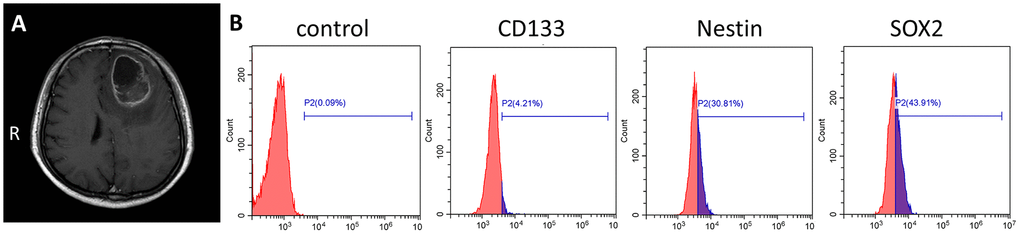
Figure 1.
Primary culture of human GSC-SU4s.
(
A
) Enhanced T1 MRI image of a 67-year-old male patient with left frontal mass. (
B
) Flow cytometric analysis of GSC markers on GSC-SU4 cells.