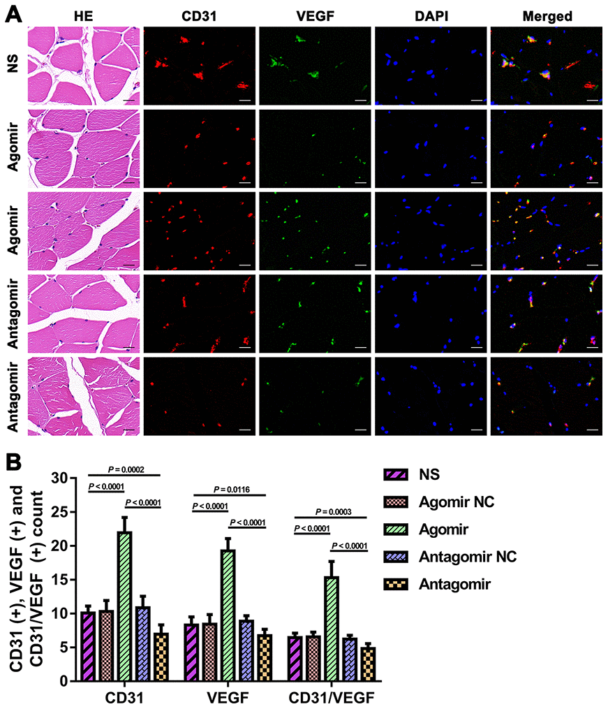
Figure 7.Expression of CD31 and VEGF in TM tissues from each group. (A) HE and immunofluorescence results showing CD31 and VEGF expression in TM tissues from each group. Bar = 20 μm. (B) Quantification of CD31(+), VEGF(+), and CD31/VEGF(+) cells in TM tissue. The data are reported as the means ± SDs. n = 8. VEGF: vascular endothelial growth factor.