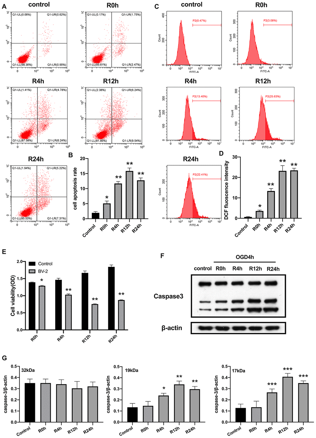
Figure 1.OGD/R induced apoptosis of BV2 microglial cells. (A, B) Flow cytometry with Annexin V/PI staining was used to assess cell apoptosis in BV2 microglial cells. (C, D) Flow cytometry was performed to measure the level of ROS in BV2 microglial cells. (E) The viability of BV2 microglial cells was determined by MTT assay. (F, G) The expression of caspase3 was quantified by western-blot analysis. All data are presented as the mean value ±SD. *p<0.05; **p<0.01, ***p<0.001, compared with control.