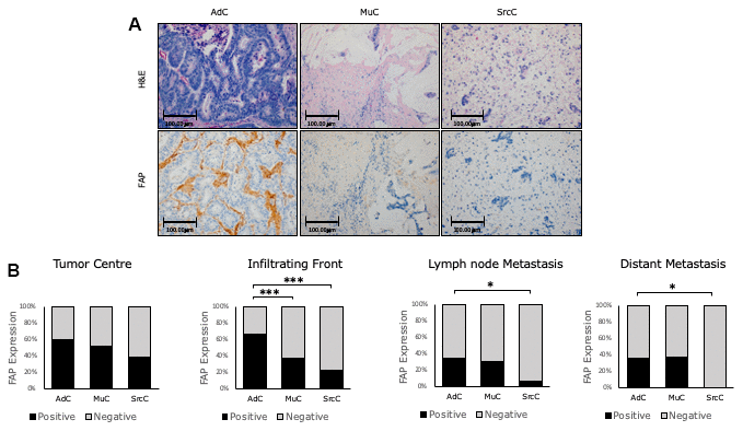
Figure 2.Immunohistochemical FAP staining according to CRC histologic subtypes. (A) Higher percentage of positive staining was observed in conventional adenocarcinoma (AdC) with respect to mucinous (MuC) and signet ring cell carcinomas (SrcC) in the infiltrating front primary tumour (x200). (B) FAP staining intensity was scored as negative or positive. The scores were quantified in each histologic subtype and statistical significance was determined by Chi-Square test (*p<0.05; ***p<0.001). H&E: Hematoxylin and Eosin. FAP: Fibroblast activation protein-α.