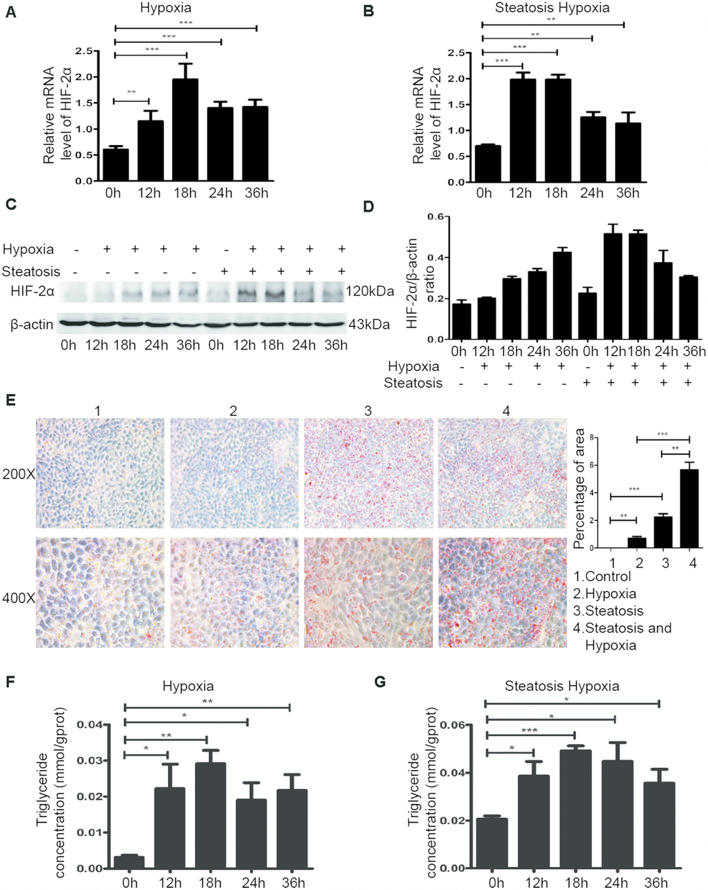
Figure 2.The hypoxic microenvironment induces lipid accumulation in HCC and steatotic HCC cells by upregulating HIF-2α. (A, B) Quantitative RT-PCR assessment of HIF-2α expression in HCC and steatotic HCC cells under hypoxic conditions. Transcription levels were normalized to those of β-actin. (C) Western blot analysis of HIF-2α expression in HCC and steatotic HCC cells under hypoxic conditions. β-Actin was used as the loading control. (D) Densitometric analyses of the band intensity ratios for HIF-2α/β-actin. (E) Oil red O staining and quantification in HCC and steatotic HCC cells with or without hypoxia treatment. (F, G) Triglyceride levels in HCC and steatotic HCC cells subjected to different durations of hypoxia.