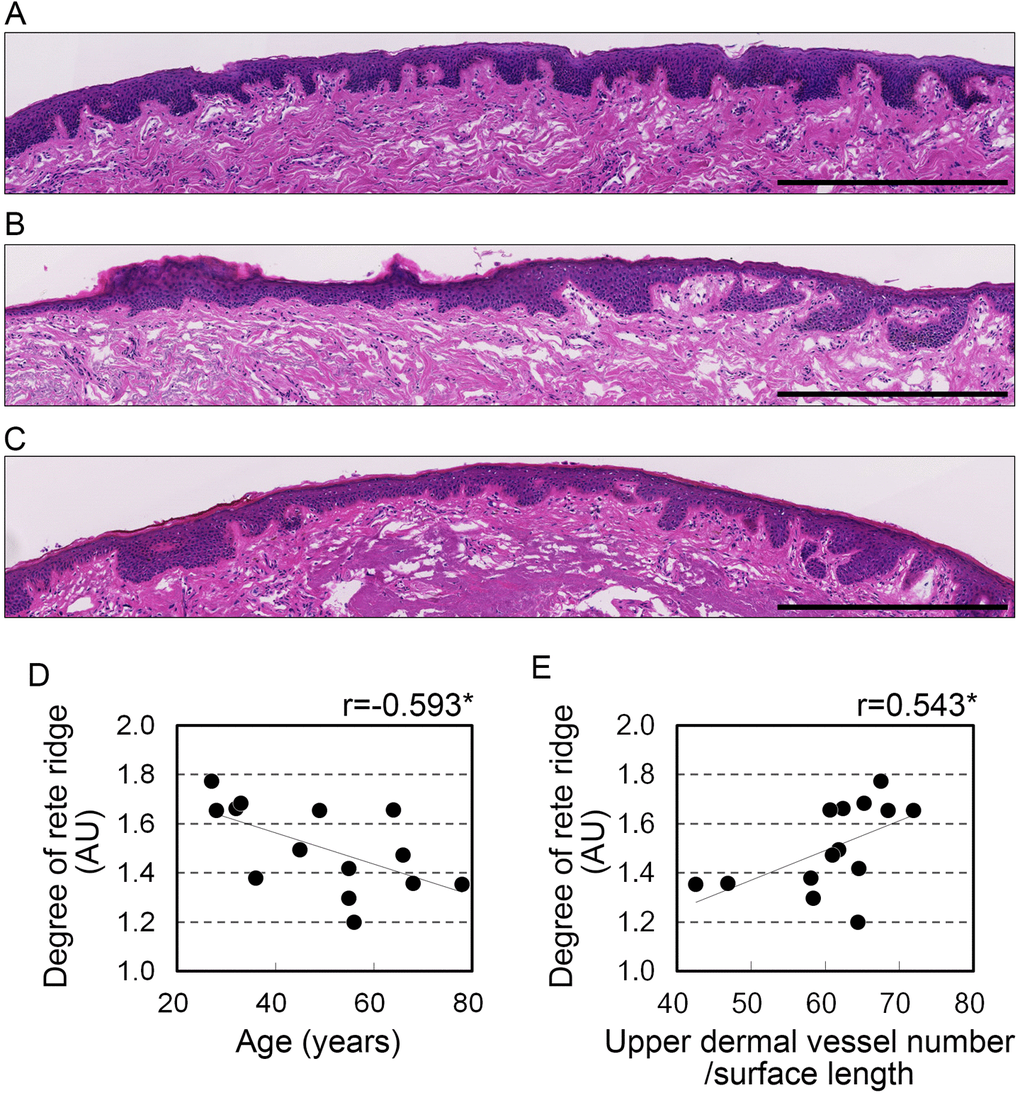
Figure 3.Development of rete ridges correlates with age and vascular condition in the upper dermis. (A-C) Representative images from hematoxylin and eosin staining in the upper dermis of 28 (A), 45 (B), and 68 (C) year old donors. Bar=500 μm. (D, E) Rete ridge elongation plotted against (D) age and (E) number of blood vessels in the upper dermis per surface length. n=14. *p<0.05; NS, not significant (Pearson's correlation test).