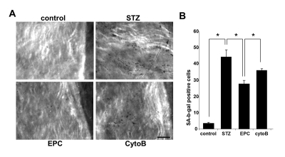
Figure 9.Senescence-associated β-galactosidase stained endothelial cells in en face aortic preparations obtained from control and STZ mice, non-treated or treated with intact EPC or with EPC preincubated with cytochalasin B (CytoB).(A) Representative images obtained using intravital microscopy. (Nikon, Melville, NY) Bars 50μm. (B)Quantitative analysis of senescence-associated β-galactosidase-stained endothelial cells. Asterisks indicate p< 0.05; n=5.