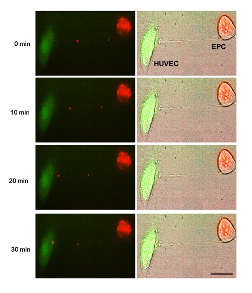
Figure 4.Representative time-lapse sequence of images illustrating a rapid transfer of lysosomes from an EPC to a stressed HUVEC.Time-lapse images of transfer of red-labeled lysosomes from EPC to stressed HUVEC labeled with CFDA SE green. Images were taken every 5 minutes. Left panels are fluorescence images and right panels are corresponding bright-field images.Bars 20μm.