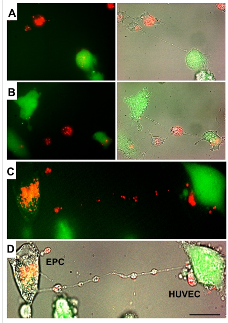
Figure 3.A gallery of images of TNT formation between HUVEC and EPC.(A, B, C and D) Typical examples of a robust transfer of lysosomes (lysotracker red) from EPC to stressed HUVEC labeled with CFDA SE green. Panels C and D depict fluorescence and bright-field images of HUVEC and EPC. Bars 20μm.