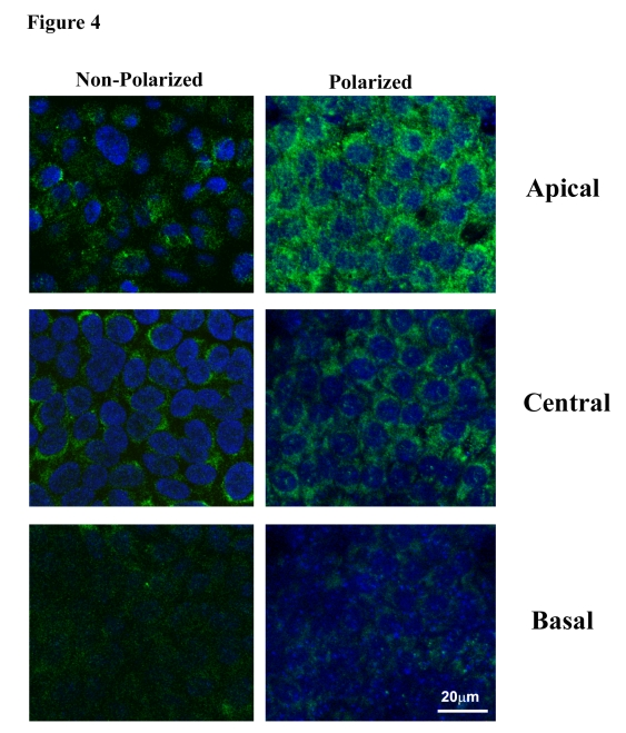
Figure 4.Distribution of PEDF in apical, central and basal regions in nonpolarized and polarized RPE cells by confocal microscopy. Staining for PEDF
is more intense in polarized RPE as compared to nonpolarized RPE. The apical
region shows much higher PEDF expression in polarized cells.