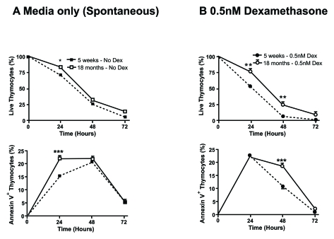
Figure 4.Aged thymocytes have increased resistance to spontaneous and dexamethasone-induced apoptosis. Spontaneous (A) and
dexamethasone (dex)-induced (B) apoptosis at 0.5nM was assessed by
flow cytometry. Graphs show the percentage of viable thymocytes defined as
Annexin V- 7AAD- (top graphs)and
those undergoing early apoptosis as Annexin V+ 7AAD-
(bottom graphs). Closed square/circle with dotted line symbolise
young thymocytes cultured in media or with the addition of 0.5nM dex
respectively. Whereas, open square/circle with solid line signify
thymocytes from 18 month old mice cultured in media or with the addition of
0.5nM dex respectively. The data revealed that there is an
age-associated increased resistance to spontaneous and dex-induced
apoptosis with a higher percentage of viable thymocytes from older mice
compared to younger mice and delayed kinetic of older thymocytes to
initiate apoptosis. Data representative of four experiments. *P<0.05;
**P<0.01.