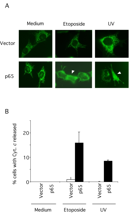
Figure 3.Cytochrome c release in p65 null cells.(A)
p65 null (vector) and reconstituted cells were treated with 10
μM etoposide or 5 mJ UV-irradiation in the presence of the caspase
inhibitor, zVAD-fmk (50 μM) for 18
hr and then fixed and stained with a specific antibody for native
cytochrome c. An Alexa Green coupled secondary antibody was
used to reveal the localization of cytochrome c. (B) Results
are expressed as percentage of cells showing cytosolic cytochrome c.