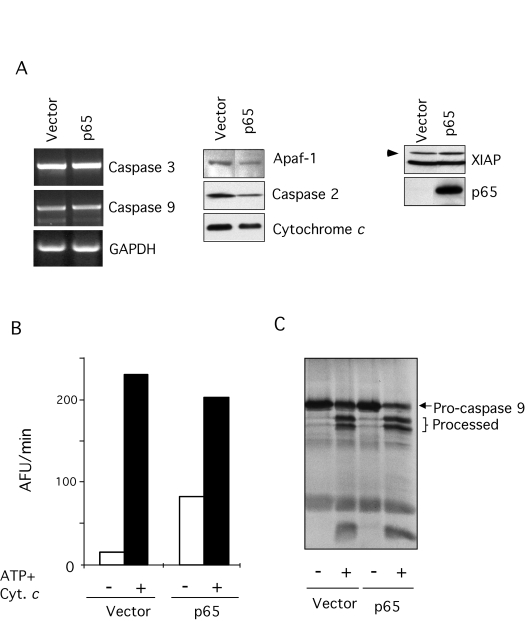
Figure 2.Characterization of the apoptotic machinery in p65 null MEFs. (A) Expression of
several key components of the apoptotic machinery in p65 null and
reconstituted cells was compared. Expression of Apaf-1, caspase-2,
cytochrome c, and XIAP was detected by immuno-blot. Expression of
caspase-3 and -9 was assessed by RT-PCR from total RNA extracted from the
cells as indicated. (B) S-100 extracts from p65 null (vector) and
reconstituted cells (p65) were incubated with 1 mM ATP and 1 μM equine cytochrome c at 37 °C for 1 hr.
Caspase activity was then assessed by cleavage (arbitrary fluorescence
units per minute [AFU/min]) of the fluorogenic substrate, Ac-DEVD-afc. (C)
caspase-9 processing by autoradiography. S-100 extracts were incubated
under the conditions described above with in vitro translated
caspase 9 and subjected to SDS-PAGE.