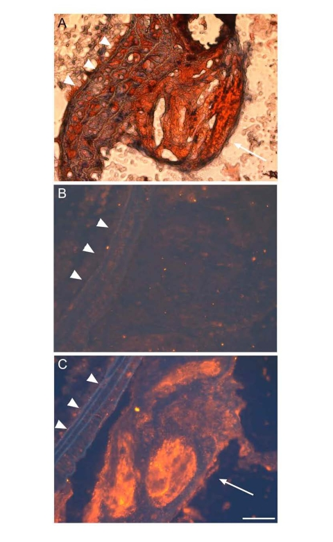
Figure 5.ApoD is localized to atherosclerotic plaques ofapoE?deficient mice.
Proximal aorta sections of male apoE knockout mice were subjected to oil red O staining (A),
and to immunohistochemistry using control rabbit IgG against bacterial β-galactosidase (B)
and rabbit anti-apoD antibody (C). The secondary antibody used in immunostaining is the donkey
anti-rabbit IgG conjugated with Cy3. ApoD was stained red in theatherosclerotic plaque (C),
as indicated by arrow. Elastic fibers ofvessels were auto-fluorescent blue,
as indicated by arrowhead. Bar = 50 μm.